|
|
|
By
the same token the author wishes to acknowledge the tremendous merits of
two electronic engineers who made the development and continuous
perfection of Instrumentation for Standardized Echography possible
through their enormous knowledge of, and skills in, the field of
diagnostic ultrasound, and through their readiness and capability to
accept and accommodate clinical necessities: (1)
Ing. Bernhard of Kretztechnik
(Zipf, Austria); with
his help the Kretz 7000 instrument of the II. Eye Department of the
University in Vienna (Austria) was modified to become the prototype for
all subsequent standardized A-scan instruments by 1964, and with his
help the author developed the first commercially available immersion
tomograph Kretztechnik 7900 S
(combined standardized A-scan and B-scan) in 1966 as well as the Kretz
7200 MA by 1970 (the only standardized A-scan unit available
throughout the 1970's and early 1980's). (2)
Jean Abascal of Biophysic Medical, later of
Biovision International [BVI] (all in Clermont-Ferrand, France),
and lastly of Quantel Medical (Bozeman, MT, USA and Clermont-Fd / Paris, France);
together with Jean Abascal the author developed the Ophthascan
S in the 1980's, the Mini-A-scan
in the late 1980's, the B-scan S in the late 1990's, and most recently, its successor, the Cine-scan
S (all with progressively increasing digital capabilities and more
numerous and powerful software programs). The
results of a collaborative international melanoma and retinoblastoma
studies undertaken for publication during the International Congress of
Ophthalmology in San Francisco in 1982 [93] demonstrate the
effectiveness of Standardized Echography performed by a great number of
echographers worldwide (all had been trained at the Universities of
Iowa, USA or Vienna, Austria): The diagnosis of
malignant melanomas of the choroid and ciliary body accomplished with
Standardized Echography by a total of 92 different examiners () in 33
different centers worldwide in a total of 1629 histological confirmed
cases carried the unique sensitivity of 99% and a specificity of 97.9%. Likewise the diagnosis
of retinoblastomas achieved by 56 different echographers in 23 different
centers worldwide in a total of 319 histological proven cases carried a
sensitivity of 98.1% and a specificity of 97.8%. Since
the mid 1970's measurements of the optic nerve [64,
66, 69, 70, 150] and the extraocular muscles [63, 64, 69,
70, 74] were added to the armamentarium of Standardized Echography
and were perfected during the following years [75,
87, 93, 94, 97,
104, 150]. The usage of the "30 degree test" [86, 87, 150]
to distinguish between solid and fluid optic nerve sheath widening and
the even more important technique of "exercising " the optic
nerve to distinguish between intracranial hypertension and optic nerve
compression [150, 173,
174, 175] were well accepted. Most reliable diagnoses of diseases
affecting the optic nerve (e.g., gliomas, meningiomas, AION, PION, optic
neuritis, optic atrophy, papilledema vs. dry optic nerve swelling,
intracranial hypertension, compressive optic neuropathy) [150], and of
diseases affecting any of the 6 extraocular muscles (e.g., Graves
orbitopathy, orbital myositis, hyperemia, hematomas, neoplasms [94, 97,
101, 104, 131,
155, 167, 176, 197, 200] were added to the widening scope of useful
diagnostic application of Standardized Echography. The
following case illustrates the applicability and great sensitivity and
accuracy of the acoustic differential criteria that have evolved over
the years for each of these ocular and orbital conditions and which can
reliably be applied because they are based on the optimal and
standardized instrument parameters and techniques of Standardized
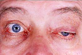
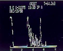
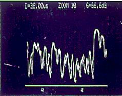
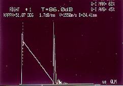
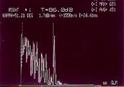
Echography: A 53-year old white male
patient had been referred to the Echography Service in the Eye
Department at the University Hospitals in Iowa City in 1990 with the
(clinical and CT-scan) diagnosis of Graves' orbitopathy. He had
undergone a previous ptosis operation OS, which was only temporally
effective. The echograms obtained with a standardized Ascan device
(Mini-A-scan) set at Tissue Sensitivity indicated medium to low
reflectivity, internal vascularity and a large angle kappa of several
tumors located within the sheaths of extraocular muscles in both orbits
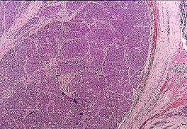
of this patient (Fig.1). On the basis of these criteria the author made
the diagnosis of metastatic carcinoid, which was later confirmed by
histopathology. This diagnosis was made
without having experienced this extremely rare kind of tumor within the
sheaths of an extraocular muscle before, on the following three
accounts: (1) the echographic findings in one case of carcinoid
metastatic to the right lacrimal fossa of a 70-year old white female
experienced 10 years earlier; her tumor echograms had shown an
3.
Phase III Since
the mid 1990's a digital revolution began to drastically change the
situation of Standardized Echography. Though some tasks of Standardized
Echography always have been easy and quick, many of the differential
diagnoses which made this method so superior to other echographic
approaches required much time, skills and experience on the part of the
echographers. Likewise the training for Standardized Echography was
lengthy and training opportunities were lacking. This situation slowed
the spread of this unique method. With
the advent of digitization and the development of powerful software
programs the situation of Standardized Echography has changed
drastically allowing for quick, easy, but still more reliable, more
objective and more accurate diagnoses to be accomplished by less trained
and less experienced echographers. This trend is just starting for
Standardized Echography and promises a bright future for this diagnostic
method. A
good example for this development is presented by the author in another
contribution to SIDUO XVIII (see Ossoinig KC: "Computer-assisted
Echographic Tissue Diagnoses: differentiation between retinal and
membranous surfaces"): The A1 sign described there had
been found to be the single most reliable and accurate acoustic
criterium for differentiating retinal detachments from dense
fibrovascular membranes. It's application was fairly quick for a well
trained and skilled echographer. With new software already contained in
the software package 2000 for the B-scan S instrument, the A1 evaluation
has become so quick and easy that untrained participants in a recent
workshop on Standardized Echography accomplished the correct diagnosis
of a retinal detachment without exception within 5 to 15 seconds. Other
features of the software package 2000 are: an automatic setting
of the Tissue Sensitivity, automatic calculations and digital readouts
of the reflectivity and sound absorption of tumor tissues. For the first
time this sound absorption can be measured and displayed in db/ mm
tissue by simply setting two electronic gates at both opposite surface
signals of the tissue or marking any section within the tissue echogram.
The associated angle kappa is measured and displayed digitally in
degrees and can be displayed schematically (Fig. 2). This
rapidly developing user-friendly automation of differential diagnoses
and measurements has begun to rejuvenate Standardized Echography
preparing it for a much wider usage in the years to come.
5.
Conclusion This
paper starts with the definition of the term "Standardized
Echography" (Introduction).
The history of the origin of Standardized Echography in early 1963, its
successes and worldwide expansion and its radical changes following the
digital revolution, which started in the early 1990 and continue with
increasing speed at the present, are narrated in Chapter 1 (Historical
Background). Chapters 2-4 describe the special characteristics of
each of the three stages in the evolution of Standardized Echography.
In
the four decades of its existence, Standardized Echography has become
the most potent and effective echographic method. Powerful software
programs developed during the past few years and being added
continuously begin to turn this sophisticated method into a quick and
easy and ever so more reliable, objective and accurate diagnostic tool
in ophthalmology, which will require much less training and experience
than in the past. 6.
Bibliography A
selection from the vast literature on Standardized Echography
(scientific papers, book chapters, and books) is presented here
including publications reporting instrumentation, techniques and
specific diagnoses for the first time or confirming previous reports
with more details or greater patient volumes, and publications
containing concise and relevant bibliographical or summarizing reviews. Scientific Papers & Book Chapters:
1. Ossoinig
KC Ultrasonic
Diagnosis of Tumors in the Eye and Orbit
- experimental and clinical examinations with the A-scan method
[German] in:
Diagnostica Ultrasonica in ophthalmologia (Proceedings of SIDUO I,
Berlin 1964, Buschmann W and Hildebrandt I, eds) pp 185-191, Math.
Naturwiss. Reihe XIV der Humboldt-Universität Berlin, 1965 2. Ossoinig
KC and Steiner H Standardization in Ultrasonic Diagnosis - a tissue model for the diagnosis of intraocular tumors
[German] in:
Diagnostica Ultrasonica in Ophthalmologia (Proceedings of SIDUO I,
Berlin 1964, Buschmann W and Hildebrandt I, eds) pp 129-133, Math.
Naturwiss. Reihe XIV der Humboldt-Universität Berlin, 1965 3.
Ossoinig KC Ultrasonic
Diagnosis in Ophthalmology [German]
9. Verhandl Österr Ophthal Ges
pp. 11-27, 1965 4.
Ossoinig KC Acoustic
Diagnosis of Ocular Tumors - Experimental and Clinical Examinations with
the A-scan Method [German]
Klin. Monatsbl. Augenheilk., 146:321-337, 1965 5.
Ossoinig KC Echography of
Intraocular Foreign Bodies [German]
Ber 67. Zus Deutsch Ophthal Ges
Heidelberg67:288-294, 1965
6. Ossoinig
KC Ultrasonic
Diagnosis of Intraocular Foreign Bodies [German] Ber. Deutsch. Ophth. Ges. 67:288-296, 1966 7.
Ossoinig KC Ultrasonic
Diagnosis of the Eye - an Aid for the Clinic (Review) in:
Ultrasonics in Ophthalmology (proceedings of Symposium Münster 1966;
Oksala A. and Gernet, H.,eds ), pp.116-133,
Karger Basel / New York 1967 8.
Ossoinig KC Evaluation
of Kinetic Properties of Echograms [German]
in: Ultrasonics in Ophthalmology (proceedings of Symposium Münster
1966; Oksala A. and Gernet, H.,eds ),
pp. 88-96, Karger Basel / New York 1967 9.
Ossoinig KC A
new Instrument for Clinical Echo-Ophthalmography [German]
Graefe's Arch. Ophthalmol. 171:312-317, 1967 10. Ossoinig KC Ultrasonic
Diagnosis of Orbital Tumors (A-scan and B-scan examinations) [German]
Graefe's Arch. Klin. Exp.Ophthal. 172:364-382, 1967 11.
Ossoinig KC and Seher K
Some Findings about the Histological Basis of Echograms [German]
in: Ultrasonics in Ophthalmology (Proceedings of Münster Symposium
1966, Oksala A
and Gernet H, eds) pp 88-96, Karger
1967 12.
Ossoinig KC and Seher K
Results in the Echography of Orbital Tumors [German]
Klin Mbl Augenheilk 151:519- 13.
Ossoinig KC and Valencak E
Echo-tomography - a Method
for the Diagnosis of Orbital Tumors [French} In: Neuro-Chir (Paris), 13:899-905, 1967 14.
Ossoinig KC The Echographic
Picture presented by Healthy Orbita (A-scan echograms) In: Diagnostica 15.
Ossoinig KC Techniques
of Echo-tomography of the Eye and Orbit [German]
In: Diagnostica Ultrasonica in Ophthalmologia (proceedings of
SIDUO II, 1967, Vanýsek, J, ed), Acta Fac. Med. Univ.Brunensis 35, pp.
117-123, Brno 1968 16.
Ossoinig KC Echography
in Ophthalmology [German]
Wien.Med. Wschr. (Proceedings sterr. Ophthalmol. Ges. 1965)
118:362-367, 1968 17.
Valencak E and Ossoinig KC
A- and B-scan Echography of
orbital A-V Fistulas [French]
in: Neuro-Chirurgie, Paris,
14:951-956, 1968 18. Böck
J and Ossoinig KC Relationship
between Histological Structures and Echograms
as the Basis for Non-traumatic Tissue Differentiation [German]
Klin Mbl Augenheilk 155:687-695
1969 19. Ossoinig
KC Routine
Ultrasonography of the Orbit
in: Ultrasonography in Ophthalmology, International Ophthalmology
Clinics 9/3:613-642, Little, Brown and Company 1969 20.
Ossoinig KC Basics, Methods,
and Results of Ultrasonography used in the Diagnosis of Intraocular Tumors in:
Ophthalmic Ultrasound (Proceedings of International Symposium in
Philadelphia 1968, Gitter KA, Keeney AH, Sarin LK, and Meyer D, eds) pp
282-293, The CV Mosby Company, St.Louis, 1969 21. Ossoinig
KC and Seher K
Ultrasonic Diagnosis of
Intraocular Foreign Bodies in:
Ophthalmic Ultrasound (Proceedings of International Symposium in
Philadelphia 1968, Gitter KA, Keeney AH, Sarin LK, and Meyer D, eds) pp.
311-320, The CV Mosby Company, St.Louis, 1969
22.
Ossoinig KC and Till P
Methods and Results of
Ultrasonography in Diagnosing Intraocular Tumors in: Ophthalmic Ultrasound (Proceedings of International
Symposium in Philadelphia 1968, Gitter KA, Keeney AH, Sarin LK, and
Meyer D, eds) pp294-300, The CV Mosby Company, St.Louis, 1969 23.
Ossoinig KC and Till P
Echo-Ophthalmography
[German] Ber
dtsch ophthal Ges 70:605-613, 1969 24.
Ossoinig KC Echography of the
Orbit - Method of Choice for the Diagnosis in Unilateral Exophthalmus
[German]
Schweiz Med Wochenschr 99:1033-1039, 1969 25. Ossoinig
KC and Valencak E Ultrasonography and
Other Diagnostic Methods: Importance in Orbital Tumors in: Ophthalmic Ultrasound (Proceedings of International
Symposium in Philadelphia 1968, Gitter KA, Keeney AH, Sarin LK, and
Meyer D, eds) pp 301-305, The CV Mosby Company, St.Louis, 1969 26. Till
P and Ossoinig KC Echographic Diagnosis of Retinoblastomas [German]
Ber Dtsch Ophthalmol Ges
69:203-209, 1969 27. Ossoinig
KC Enophthalmus
- a Sign of an orbital Tumor[German]
Ber. Deutsch Ophthal Ges Heidelberg 1969, JF Bergmann Verlag,
München, 1970 28. Gerstner
R. and Ossoinig KC A New High-frequency Ultrasonic Instrument for the Diagnosis and
Differential Diagnosis of abnormal
Tissues [German] in:
Ultrasonographia Medica (Proceedings of 1st World Congress on
Ultrasonic Diagnostics in Medicine and SIDUO III, Vienna 1969 (Böck J
and Ossoinig KC, eds) Vol I, pp. 55-60, published by Wien. Med. Akad.,
Wien 1971. 29. Ossoinig
KC Basics
of Clinical Echo-Ophthalmography. Part IV: Clinical Standardization of
Instrument and Examination Techniques [German] in: Ultrasonographia Medica (Proceedings of 1st
World Congress on Ultrasonic Diagnostics in Medicine and SIDUO III,
Vienna 1969 (Böck J and Ossoinig KC, eds) Vol II, pp. 83-118, published
by Wien. Med. Akad., Wien 1971 30. Ossoinig
KC Ultrasonic
Diagnosis of Orbital Vascular Lesions [German] Klin Mbl Augenheilk 158:526-533 1971 31. Ossoinig
KC Echo-Orbitography
- a reliable method
for the Differential Diagnosis of Endocrine Exophthalmus
in: Further Advances in Thyroid Research (Proc of Vienna
Conference on Endocrine Disorders 1968, Fellinger K and Höfer R, eds)
pp 871-877, Verlag Wiener
Med Akad, Vienna 1971
32. Till
P Echography
of Retrobulbar Hematomas [German] Klin
Mbl. Augenheilk 158:723, 1971 33. Till
P and Neumann A Reliability of Biological Standards [German] ]
in: Ultrasonographia Medica (Proceedings of 1st
World Congress on Ultrasonic Diagnostics in Medicine and SIDUO III,
Vienna 1969 (Böck J and Ossoinig KC, eds) Vol II, pp. 119-124,
published by Wien. Med. Akad., Wien 1971 34.
Bronson NR Development
of a Simple B-scan Ultrasonoscope. Trans. Amer. Ophthal. Soc. 70:
365-408, 1972 35.
Ossoinig KC Clinical Echo-Ophthalmography
in: Current Concepts in Ophthalmology (Blodi FC, ed)
8:101-130,
The CV Mosby Company, 1972 36.
Ossoinig KC The Clinical
Value of Echography in Traumatic Retinal Detachments [German] Mod Probl Ophthal
10:437-442, 1972 37.
Till P and Ossoinig KC
Echography in the
Evaluation of periorbital Carcinomas invading the Orbit [German]
Msch
Ohrh pp 442-448, 1972 38.
Till P and Ossoinig KC
Echographic Findings in
Retinopathia Proliferans Klin
Mbl Augenh 161: 297-301,
1972 39.
Freyler H
Echography in Hemorrhagic
Choroidal Detachments [German] ] in:
Diagnostica Ultrasonica in Ophthalmologia (Proceedings of SIDUO IV,
Paris 1971; Massin M. and Poujol J., eds) pp. 189-193,
published by Centre National d'Ophtalmologie des Quinze-Vingts,
Paris 1973 40.
Ossoinig KC A
New Instrument for Clinical Echo-Ophthalmography (proposals for the
standardization of important instrument parameters) [German]
in: Diagnostica Ultrasonica in Ophthalmologia (Proceedings of
SIDUO IV, Paris 1971; Massin M. and Poujol J., eds) pp. 131-137,
published by Centre National d'Ophtalmologie des Quinze-Vingts,
Paris 1973 41.
Ossoinig KC Discussion of
"Reliability of Ocular Tumor Diagnosis with Ultrasound" by
Coleman DJ Trans
Amer
Acad Ophthalmol Otolaryngol 77/5 OP: 684-686, 1973 42.
Till P and Abel JO
Results of Echography in
Cases with Detached Retina [German]
in: Diagnostica Ultrasonica in Ophthalmologia (Proceedings of
SIDUO IV, Paris 1971; Massin M. and Poujol J., eds) pp.195-198,
published by Centre National d'Ophtalmologie des Quinze-Vingts,
Paris 1973 43. Ossoinig
KC Preoperative
Differential Diagnosis of Tumors with Echography. I. Physical Principles
and Morphological Background of Tissue Echograms
In: Current Concepts in Ophthalmology (Blodi FC ed)
4:264-280. CV Mosby, St.Louis 1974 44. Ossoinig
KC Preoperative
Differential Diagnosis of Tumors with Echography. II. Instrumentation
and Examination Techniques In:
Current Concepts in Ophthalmology (Blodi FC ed) 4:280-296. CV Mosby,
St.Louis 1974 45. Ossoinig
KC Quantitative
Echography - An Important Aid for the Acoustic Differentiation of
Tissues in:
Ultrasonics in Medicine (Proceedings of 2nd World Congress on
Ultrasonics in Medicine, Rotterdam 1973, de Vlieger M, White DN and
McCready VR, eds) pp 49-54, Excerpta Medica, Amsterdam 1974 46.
Ossoinig KC Quantitative
Echography - the basis of tissue differentiation J Clinical Ultrasound 2/1: 33-46, 1974
47. Ossoinig
KC and Blodi FC Preoperative Differential Diagnosis of Tumors with Echography.
III Diagnosis of Intraocular Tumors
In: Current Concepts in Ophthalmology (Blodi FC ed)
4:296-313. CV Mosby, St.Louis 1974 48. Ossoinig
KC and Blodi FC Preoperative Differential Diagnosis of Tumors with Echography . IV.
Diagnosis of Orbital Tumors In:
Current Concepts in Ophthalmology (Blodi FC ed) 4:313-341. CV Mosby,
St.Louis 1974 49. Till
P and Lessel MR Doppler-Sonography
and Echo-Ophthalmography off Orbital Vascular Processes
in: SIDUO Round Table 1974 (Massin M and Poujol J, eds) pp
51-58, Centre Nat d'Ophtalmol des Quinze-Vingts, Paris 1974 50. Freyler
H and Arnfelser H Relation between Histological Structure and Ultrasonogram in Malignant
Melanoma of the Choroid in:
Ultrasonography in Ophthalmology
(Proceedings of SIDUO V, Francois J and Goes F, eds) pp
163-171, S Karger 1975 51. Ossoinig
KC A-scan
Echography and Orbital Disease
in: Modern Problems in Ophthalmology (Proceedings 2nd
Int Symposium on Orbital Disorders, Amsterdam 1973 (Streiff EB,
ed) Vol 14, pp 203-235, Karger 1975 52. Ossoinig
KC, Bigar F and Kaefring S Malignant
Melanoma of the Choroidand Ciliary Body. A differential diagnosis in
Clinical Echography in: Ultrasonography in Ophthalmology (Proceedings of SIDUO
V, Francois J and Goes F, eds) pp141-154,
S Karger 1975 53. Ossoinig
KC, Bigar F, Kaefring S and McNutt L
Echographic Detection and
Localization of BB Shots in the Eye and Orbit
in: Ultrasonography in Ophthalmology
(Proceedings of SIDUO V, Francois J and Goes F, eds) pp
109-118, S Karger 1975 54. Ossoinig
KC, Keenan TP and Bigar F Cavernous
Hemangioma of the Orbit in:
Ultrasonography in Ophthalmology
(Proceedings of SIDUO V, Francois J and Goes F, eds) pp
236-244, S Karger 1975 55. Ossoinig
KC and Till P Ten-year Study on Clinical Echography in Orbital Disease
Bibl Ophthalmol 83:200 1975 56. Ossoinig
KC and Till P Clinical Echography of Tumors in the Eye and Orbit [German]
in: Krebsbehandlung als Interdisziplinäre Aufgabe, Kärcher
KH ed, pp 249-270, Springer Verlag 1975 57. Till
P Echography
in Rhinogenic Orbital Conditions in:
Modern Problems in Ophthalmology: Orbital Disorders (Bleeker GM et al,
eds) Vol 14, p 273, Karger (Basel) 1975 58. Till
P and Ossoinig K Ten-year Study on Clinical Echography in Intraocular Disease
Bibl Ophthalmol 83:49 1975 59. Wüstenberg
L and Kohlhase R Echographic Findings in Unilateral Exophthalmus [German]
in: Ultrasonography in Ophthalmology
(Proceedings of SIDUO V, Francois J and Goes F, eds) pp
217-223, S Karger 1975 60. Ossoinig
KC Clinical
Echography of Orbital Tumors
in: proc Sympos Amer Soc Plast Reconstr Surgeons, Dallas
1974, Tessier P, Callahan A, Mustarde JC and Salyer KE, eds; 12:307-315,
CV Mosby 1976 61. Till
P Solid
Tissue Model for the Standardization of the Echo-Ophthalmograph 7200 MA
(Kretztechnik) Docum
Ophthalmol 41:205 1976 62. Hodes
BC Standardized A-scan Echographic
Diagnosis of Choroidal Malignant Melanomas
Arch.Ophthal 95:593-597,
1977 63. McNutt
LC, Kaefring SL, and Ossoinig KC Echographic
Measurement of Extraocular Muscles
in: Ultrasound in Medicine, Vol 3A (Proceedings of 1st
Triennial Meeting [2nd World Congress] of the World Federation for
Ultrasound in Medicine and Biology and SIDUO VI, San Francisco, 1976,
White D. and Brown RE, eds) pp. 927-932,
Plenum Press, New York 1977 64. Ossoinig
KC Echography
of the Eye, Orbit and Periorbital Region
in: Orbit Roentgenology (Arger PH, ed), 224-269, John Wiley
and Sons, New York 1977 65. Ossoinig
KC Special
Echographic Techniques for the Evaluation of Ocular Foreign Bodies
[German] in:
proc of "Intraokulärer Fremdkörper und Metallose", Symp DOG
Köln 1976, Neubauer H, 66.
Ossoinig KC, Kaefring SL, McNutt LC, and Weinstock
SJ Echographic
Measurement of the Optic Nerve in:
Ultrasound in Medicine, Vol 3A (Proceedings of 1st Triennial Meeting
[2nd World Congress] of the World Federation for Ultrasound in Medicine
and Biology and SIDUO VI, San Francisco, 1976,
White D. and Brown RE, eds) pp. 1065-1066,
Plenum Press, New York 1977 67.
Ossoinig KC, Frazier S, Watzke RC and Diamond JG
Combined A-scan and B-scan Echography as a Diagnostic
Aid for Vitreoretinal Surgery
in: New and Controversial Aspects of Vitreoretinal
Surgery, Houston 1975, McPherson
A, ed; pp106-125, CV Mosby 1977 68.
Till P and Ossoinig KC
First Experiences with a
Solid Tissue Model for the Standardization of A- and B-scan Instruments in Tissue
Diagnosis in: Ultrasound in Medicine (Proceedings of SIDUO
VI, 1976, White D and Brown RE,
eds) Vol 3B, pp 2167-2174, Plenum Publishing Corp, 1977 69. Ossoinig
KC Echography
of Orbital Disorders in:
Clinical Handbook of Ultrasound (M de Vlieger, ed) pp 881-904, John
Wiley and Sons, Inc. (New York) 1978 70. Ossoinig
KC The
Role of Clinical Echography in Modern Diagnosis of Periorbital and
Orbital Lesions in:
Proceedings of 3rd Int Symposium on Orbital Disorders,
Amsterdam 1977 Bleeker GM, ed) pp 496-540, Dr.W.Junk Publishers, The
Hague 1978 71. Fuller
DG, Snyder WB, Hutton WL and Vaiser A
Ultrasonographic Features
of Choroidal Malignant Melanomas
Arch Ophthalmol 97:1465-1472, 1979 72. Hauff
W and Till P
Echographic Findings in
Orbital Mucoceles in: Diagnostica Ultrasonica in Ophthalmologia (Proceedings of SIDUO
VII, Gernet H., ed) pp 151-154, R.A.Remy - Verlag, Münster 1979
73. Mazzeo
V and Scorrano R Our Echographic Experience about Choroidal Melanomas
in: Diagnostica Ultrasonica in Ophthalmologia (Proceedings of SIDUO VII, Gernet H., ed) pp 119-122,
R.A.Remy - Verlag, Münster 1979 74. Ossoinig
KC The
Technique of Measuring Extraocular Muscles
in: Diagnostica Ultrasonica in Ophthalmologia (Proceedings of SIDUO
VII, Gernet H., ed) pp 166-172, R.A.Remy - Verlag, Münster 1979 75. Ossoinig
KC Standardized
Echography: Basic Principles, Clinical Applications, and Results
In: Ophthalmic Ultrasonography: Comparative Techniques (RL Dallow,
editor), International Ophthalmology Clinics 19:No.4, pp.127-210, 1979 76. Scott
W, Fabre J and Ossoinig KC Congenital Mucocele of the Lacrimal Sac Arch Ophthal 9/1, pp 1656-1658 1979 77. Till
P Echography
in Intraocular Tumors in:
Diagnostica Ultrasonica in Ophthalmologia
(Proceedings of SIDUO VII, Gernet H., ed) R.A.Remy - Verlag,
Münster 1979 78. Dagher
G, Anderson R, Ossoinig KC and Baker J
Adenoid Cystic Carcinoma of
the Lacrimal Gland in a Child
Arch Ophthal 98:1098-1100, 1980 79. Ossoinig
KC and Blodi FC The Role of Echography in the Diagnosis of Orbital Disorders
Palestra Oftalm Panamericana 4:12-19, 1980 80.
Till P Testing
of Ultrasonic Probes and their Suitability for Tissue Differentiation
with the help of the Solid Tissue
Model [German] Klin Mbl Augenheilk 176: 337-340, 1980 81. Gallenga
PE, Dalia T, Bellone G, Cennamo G, Mazzeo V and Rossi A The Role of
Echography in the Conservative
Treatment of endobulbar Tumors
in:
Ultrasonography in Ophthalmology
(Documenta Ophthalmologica Proceedings Series 29, Thijssen JM
and Verbeek AM, eds) pp 117-122, Dr.
W. Junk Publishers 1981 82. Hauff
W and Till P Echography in
Unusual Orbital Complications of Parasinusal Diseases
in: Ultrasonography in Ophthalmology (Documenta
Ophthalmologica Proceedings Series 29, Thijssen JM and Verbeek AM, eds)
pp 317-322, Dr. W. Junk
Publishers 1981 83. Nishimoto
Y, Baba Y, Shibata H and Sawada A
Combination of Echography
with Coronal CT in the Diagnosis of Orbital Disorders
in: Ultrasonography in Ophthalmology
(Documenta Ophthalmologica Proceedings Series 29, Thijssen JM
and Verbeek AM, eds) pp 301-316, Dr.
W. Junk Publishers 1981 84. Ossoinig
KC Echographic
Differentiation of Vascular Tumors in the Orbit in: Ultrasonography in Ophthalmology (Documenta
Ophthalmologica Proceedings Series 29, Thijssen JM and Verbeek AM, eds)
pp 283-291, Dr. W. Junk
Publishers 1981 85.
Ossoinig KC The Significance
of the S-shaped Amplifier characteristics in Echographic Tissue
Diagnosis in:
Ultrasonography in Ophthalmology
(Documenta Ophthalmologica Proceedings Series 29, Thijssen JM
and Verbeek
AM, eds) pp 441-443, Dr.
W. Junk Publishers 1981 86.
Ossoinig KC The
Significance of Standardized Echography for Neuro-ophthalmology
in: "Aktuelle Ophthalmologische Probleme (Remke H, ed)
pp 61-75, Ferdinand Enke Verlag, Stuttgart 1981 87. Ossoinig
KC, Cennamo G and Byrne SF Echographic
Differential Diagnosis of Optic-Nerve Lesions
in: Ultrasonography
in Ophthalmology
(Documenta Ophthalmologica Proceedings Series 29, Thijssen JM
and Verbeek AM, eds) pp 327-332, Dr.
W. Junk Publishers 1981 88. Ossoinig
KC, Cennamo G, Green RL and Weyer, NL
Echographic Results in the
Diagnosis of Retinoblastoma in:
Ultrasonography in Ophthalmology
(Documenta Ophthalmologica Proceedings Series 29, Thijssen JM
and Verbeek AM, eds) pp 103-107, Dr. W. Junk Publishers 1981 89. Till
P and Hauff W Differential
Diagnostic Results of Clinical Echography in Intraocular Tumors
in: Ultrasonography in Ophthalmology
(Documenta Ophthalmologica Proceedings Series 29, Thijssen JM
and Verbeek AM, eds) pp 91-95, Dr. W. Junk Publishers 1981
90. Till
P and Hauff W Differential
Diagnostic Results of Clinical Echography in Orbital Tumors
in: Ultrasonography in Ophthalmology
(Documenta Ophthalmologica Proceedings Series 29, Thijssen JM
and Verbeek AM, eds) pp 277-282, Dr.
W. Junk Publishers 1981
91. Till
P and Scheiber V Reliability and Accuracy of TM (Tissue Model) for Calibration of
Standardized A-scan Instrumentation
in: Ultrasonography
in Ophthalmology
(Documenta Ophthalmologica Proceedings Series 29, Thijssen JM
and Verbeek AM, eds) pp 457-461, Dr.
W. Junk Publishers 1981 92.
Divine RD, Anderson RL and Ossoinig KC
Metastatic Carcinoid
Unresponsive to Radiation Therapy presenting asa Lacrimal Fossa Mass Ophthalmology 89:516-520, 1982 93. Ossoinig
KC Advances
in Diagnostic Ultrasound in:
Acta:XXIV International Congress of Ophthalmology, San Francisco1982
(Paul Henkind, ed.), J.B.Lippincott Comp., Vol1, pp.89-114, 1982.
94. Ossoinig
KC A
New Echographic Sign for a Reliable Differential Diagnosis of Graves'
Orbitopathy [German] Klin
Mbl Augenheilk 180:189-197,
1982 95. Phelps
C, Thompson S and Ossoinig KC
The Diagnosis and Prognosis
of Atypical Carotid-Cavernous Fistula (Red-Eyed Shunt Syndrome) AJO 93:423-436, 1982 96. Ossoinig
KC and Harrie RP Diagnosis of Intraocular Tumors with Standardized Echography
in: Intraocular Tumors (Proceedings of International
Symposium in Schwerin, 1981, Lommatzsch PK and Blodi FC, eds), pp.
154-175, Akademie Verlag Berlin, 1983 97. Ossoinig
KC and Hasenfratz G The Role of Standardized Echography in the Diagnosis and Treatment of
Orbital Myositis [German] Fortschr
Ophthalmol 80:475-481 1983 98. Byrne
SF Standardized
Echography in the Diagnosis of Hemangioendothelioma in: Ophthalmic Ultrasonography (Documenta
Ophthalmologica Proceedings Series 38, Hillman JS and Le May MM, eds) pp
347-356, Dr. W. Junk Publishers (The Hague/Boston/Lancaster) 1984 99. Hasenfratz
G and Ossoinig KC The
Diagnosis of Orbital Mucoceles and Pyoceles with Standardized Echography
in: Ophthalmic Ultrasonography (Documenta Ophthalmologica Proceedings Series 38, Hillman
JS and Le May MM, eds) pp 407-415, Dr. W. Junk Publishers (The
Hague/Boston/Lancaster) 1984
1100. Hauff
W and Till P Echography in
Carotid-Cavernous Fistulas in:
Ophthalmic Ultrasonography
(Documenta Ophthalmologica Proceedings Series 38, Hillman JS
and Le May MM, eds) pp 399-405, Dr. W. Junk Publishers (The
Hague/Boston/Lancaster) 1984 1101. Ossoinig
KC Ultrasonic
Diagnosis of Graves' orbitopathy in:
The Eye and Orbit in Thyroid Disease (Gorman CA, Waller RR, and Dyer JA,
eds) Raven Press, New York, pp.185-211, 1984
1102. Ossoinig
KC Echographic
Detection and Classification of Posterior Hyphemas Ophthalmologica 189:2-11, 1984 103. Ossoinig
KC How
to obtain Maximum Measuring Accuracies with Standardized A-scan
in: Ophthalmic Ultrasonography
(Documenta Ophthalmologica Proceedings Series 38, Hillman JS
and Le May MM, eds) pp 197-216, Dr. W. Junk Publishers (The
Hague/Boston/Lancaster) 1984 104. Ossoinig
KC and Hermsen VM Myositis of Extraocular Muscles diagnosed with Standardized Echography
in: Ophthalmic
Ultrasonography
(Documenta Ophthalmologica Proceedings Series 38, Hillman JS
and Le May MM, eds) pp 381-392, Dr. W. Junk Publishers (The
Hague/Boston/Lancaster) 1984 105. Ossoinig
KC and Tamayo G Ultrasonic Differential Diagnosis of Lacrimal-Gland Tumors
in: Supplement to J. of Ultrasound in Medicine, Vol 4, No 10,
pp 52- , 1985
106. Byrne
SF and Byrne B Differential Diagnosis of orbital Neurolemmoma (schwannoma) with
Standardized Echography
in: Ophthalmic Echography (Documenta Ophthalmologica
Proceedings Series 48, Ossoinig KC, ed) pp 483-492, Martinus Nijhoff /
Dr. Junk Publishers (Dordrecht/Boston/Lancaster) 1987
107. Byrne
SF and Hughes JR Orbital Dermoid Cysts
in: Ophthalmic Echography (Documenta Ophthalmologica
Proceedings Series 48, Ossoinig KC, ed) pp 465-475, Martinus Nijhoff /
Dr. Junk Publishers (Dordrecht/Boston/Lancaster) 1987 108. Dixon
PA, Abrams GW and Caya JG Acoustic analysis of the cytologic structure of malignant melanomas with
Standardized Echography
in: Ophthalmic Echography (Documenta Ophthalmologica
Proceedings Series 48, Ossoinig KC, ed) pp 347-356, Martinus Nijhoff /
Dr. Junk Publishers (Dordrecht/Boston/Lancaster) 1987 109. Fishman
M, Kerman B and Foxman S Intraocular
Cysticercosis: migratory
in: Ophthalmic Echography (Documenta Ophthalmologica
Proceedings Series 48, Ossoinig KC, ed) pp 285-287, Martinus Nijhoff /
Dr. Junk Publishers (Dordrecht/Boston/Lancaster) 1987 110. Goes
F Ultrasonographic
and Clinical Characteristics of Orbital Pseudotumors in: Ophthalmic Echography (Documenta
Ophthalmologica Proceedings Series 48, Ossoinig KC, ed) pp 499-507,
Martinus Nijhoff / Dr. Junk Publishers (Dordrecht/Boston/Lancaster) 1987
111. Green
RL The
Echographic Evaluation of Spontaneous Vitreous Hemorrhage
in: Ophthalmic Echography (Documenta Ophthalmologica
Proceedings Series 48, Ossoinig KC, ed) pp 233-238, Martinus Nijhoff /
Dr. Junk Publishers (Dordrecht/Boston/Lancaster) 1987
112. Green
RL Echographic
Diagnosis of Posterior Scleritis
in: Ophthalmic Echography (Documenta Ophthalmologica
Proceedings Series 48, Ossoinig KC, ed) pp 515-519, Martinus Nijhoff /
Dr. Junk Publishers (Dordrecht/Boston/Lancaster) 1987 113. Harrie
RP Standardized
Echography of the Orbit (review)
in: Ophthalmic Echography (Documenta Ophthalmologica
Proceedings Series 48, Ossoinig KC, ed) pp 445-451, Martinus Nijhoff /
Dr. Junk Publishers (Dordrecht/Boston/Lancaster) 1987
114. Hasenfratz
G Standardized
Echography in Graves' disease
in: Ophthalmic
Echography (Documenta Ophthalmologica Proceedings Series 48, Ossoinig
KC, ed) pp 557-564, Martinus Nijhoff / Dr. Junk Publishers (Dordrecht/Boston/Lancaster)
1987 115. Hughes
JR and Byrne SF Detection of Posterior Ruptures in Opaque Media
in: Ophthalmic Echography (Documenta Ophthalmologica
Proceedings Series 48, Ossoinig KC, ed) pp 333-342, Martinus Nijhoff /
Dr. Junk Publishers (Dordrecht/Boston/Lancaster) 1987 116. Kerman
BM and Fishman ML Non-melanomatous Collar-button Tumors in: Ophthalmic Echography (Documenta
Ophthalmologica Proceedings Series 48, Ossoinig KC, ed) pp 413-416,
Martinus Nijhoff / Dr. Junk Publishers (Dordrecht/Boston/Lancaster) 1987
117. Ossoinig
KC Early
Detection of Compressive Optic Neuropathy in Graves' disease with
Standardized A-scan in:
Ophthalmic Echography (Documenta Ophthalmologica Proceedings Series 48,
Ossoinig KC, ed) p 569, Martinus Nijhoff / Dr. Junk Publishers (Dordrecht/Boston/Lancaster)
1987 118. Ossoinig
KC and Cody K How to differentiate intraocular air bubbles from intraocular foreign
bodies using Standardized Echography
in: Ophthalmic Echography (Documenta Ophthalmologica
Proceedings Series 48, Ossoinig KC, ed) pp 343-344, Martinus Nijhoff /
Dr. Junk Publishers (Dordrecht/Boston/Lancaster) 1987 119. Ossoinig
KC, Frieling E, Tamburrelli C and Warner L
Superior Ophthalmic Vein
Thrombosis - an echographic diagnosis
in: Ophthalmic Echography (Documenta Ophthalmologica
Proceedings Series 48, Ossoinig KC, ed) pp
528-537, Martinus Nijhoff / Dr. Junk Publishers (Dordrecht/Boston/Lancaster)
1987
120. Ossoinig
KC, Islas G, Tamayo GE and Tamburrelli C
Detached Retina versus
dense Fibrovascular Membrane. Standardized A-scan and B-scan criteria in: Ophthalmic Echography (Documenta
Ophthalmologica Proceedings Series 48, Ossoinig KC, ed) pp 275-284,
Martinus Nijhoff / Dr. Junk Publishers (Dordrecht/Boston/Lancaster) 1987 121. Ossoinig
KC and Itani KM The Echographic Diagnosis of Non-Calcified Retinoblastoma
in: Ophthalmic Echography (Documenta Ophthalmologica
Proceedings Series 48, Ossoinig KC, ed) p 444, Martinus Nijhoff / Dr.
Junk Publishers (Dordrecht/Boston/Lancaster) 1987
122. Ossoinig,
KC, Reshef DS, Weingeist TA, Folk JC and Packer AJ Echogrphic
Findings in Terson's Syndrome
in: Ophthalmic Echography (Documenta Ophthalmologica
Proceedings Series 48, Ossoinig KC, ed) pp 247-256, Martinus Nijhoff /
Dr. Junk Publishers (Dordrecht/Boston/Lancaster) 1987
123. Ossoinig
KC and Tamburrelli C Orbital Aerocele in:
Ophthalmic Echography (Documenta Ophthalmologica Proceedings Series 48,
Ossoinig KC, ed) p 497, Martinus Nijhoff / Dr. Junk Publishers (Dordrecht/Boston/Lancaster)
1987 124. Reshef
DS, Ossoinig KC and Nerad JA
Diagnosis and
Intraoperative Localization of a Deep Orbital Organic Foreign Body
Orbit: Vol 6/1, pp 3-15,
1987 125. Rochels
R, Scherer U, Nover A and Lieb W
Echographical Diagnosis of
Lacrimal Sac Tumors in:
Ophthalmic Echography (Documenta Ophthalmologica Proceedings Series 48,
Ossoinig KC, ed) pp 545-552, Martinus Nijhoff / Dr. Junk Publishers (Dordrecht/Boston/Lancaster)
1987 126. Sawada
A, Masuyama Y, Kodama Y and Hayashida T
Combined Echography and
Fluorophotometry in the Detection od Vitreous Hemorrhage
in: Ophthalmic Echography (Documenta Ophthalmologica
Proceedings Series 48, Ossoinig KC, ed) pp 239-246, Martinus Nijhoff /
Dr. Junk Publishers (Dordrecht/Boston/Lancaster) 1987
127. Shammas
HJ, Boyer DS and Miller JB Ultrasound
Characteristics of posterior Uveal Melanomas treated with Cobalt Plaque
Radiotherapy in:
Ophthalmic Echography (Documenta Ophthalmologica Proceedings Series 48,
Ossoinig KC, ed) pp 379-383, Martinus Nijhoff / Dr. Junk Publishers (Dordrecht/Boston/Lancaster)
1987 128. Skalka
HW Ultrasound
Diagnoses of Orbital Masses and Intraocular Tumors in: Ophthalmic Echography (Documenta
Ophthalmologica Proceedings Series 48, Ossoinig KC, ed) pp 463-464,
Martinus Nijhoff / Dr. Junk Publishers (Dordrecht/Boston/Lancaster) 1987
129. Stone
RD Regression
patterns of choroidal malignant melanoma: Standardized Echography
(A-mode) and immersion tomography (B-mode). A comparative study in: Ophthalmic Echography (Documenta
Ophthalmologica Proceedings Series 48, Ossoinig KC, ed) pp 233-238,
Martinus Nijhoff / Dr. Junk Publishers (Dordrecht/Boston/Lancaster) 1987
130. Verbeek
AM Uveal
Melanomas before and after Ruthenium Application Therapy in:
Ophthalmic Echography (Documenta Ophthalmologica Proceedings Series 48,
Ossoinig KC, ed) pp. 385-389, Martinus Nijhoff / Dr. Junk Publishers (Dordrecht/Boston/Lancaster)
1987 131.
Ossoinig KC The Role of
Standardized Ophthalmic Echography in the Management of Graves'
opthalmopathy in:
Graves' Ophthalmopathy (Pickardt CR and Boergen KP, eds) pp
28-37, Developments in ophthalmology, Karger
1989 132. Badia
JA, Degregori W, Iribarren G and Sampaolesi R
Reliability of Standardized
Ultrasound in Pre-operative
Diagnosis for Vitreous Surgery in Diabetic Patients
in: Ultrasonography in Ophthalmology 12 (Documenta Ophthalmologica Proceedings Series 53, Sampaolesi R, ed)
pp. 233-237, Kluwer Academic Publishers 1990
133. Cennamo
G, Tranfa F and Bonavolonta G
Lesions of the Lacrimal
Fossa: a retrospective echographic study
in: Ultrasonography in Ophthalmology 12
(Documenta Ophthalmologica Proceedings Series 53, Sampaolesi R, ed)
pp. 63-78, Kluwer Academic Publishers 1990 134. Lombardi
A, Croxatto JO and Zambrano A
Diffuse Lymphoid
Infiltration of the Uvea and Periocular Tissues
in: Ultrasonography in Ophthalmology 12
(Documenta Ophthalmologica Proceedings Series 53, Sampaolesi R, ed)
pp. 427-437, Kluwer Academic Publishers 1990
135.
Mazzeo V, Ravalli L and Perri P
Echographic Findings in
Lymphoid Hyperplasia of the Choroid in: Ultrasonography in Ophthalmology 12 (Documenta Ophthalmologica Proceedings Series 53, Sampaolesi R, ed)
pp. 419-425, Kluwer Academic Publishers 1990 136. Ossoinig
KC and Lohmeyer M Choroidal Nevi: diagnosis with Standardized Echography
in: Ultrasonography in Ophthalmology 12
(Documenta Ophthalmologica Proceedings Series 53, Sampaolesi R, ed)
pp. 173-180, Kluwer Academic Publishers 1990
137. Schutterman
J
A case with a macular
granuloma seropositive for Toxocara canis examined with Standardized
Echography in:
Ultrasonography in Ophthalmology 12
(Documenta Ophthalmologica Proceedings Series 53, Sampaolesi R, ed)
pp. 261-265, Kluwer Academic Publishers 1990
138.
Zárate JO and Sampaolesi R
Morphological Parameters in
Intraocular Tumors taking part on Echographic Tracings
in: Ultrasonography in Ophthalmology 12
(Documenta Ophthalmologica Proceedings Series 53, Sampaolesi R, ed)
pp. 281-292, Kluwer Academic Publishers 1990
139. Ossoinig
KC Detection of
Wooden Foreign Bodies . Letter to the Editor, Ophthalmology 98/3
p 274, 1991 140. Atta
HR Standardized
Echography in a Case of
Orbital Varix in:
Ophthalmic Echography 13
(Documenta Ophthalmologica Proceedings Series 55, Till P, ed)
pp. 199-203, Kluwer Academic Publishers 1993
141. Cennamo
G, Loffredo A, Rosa N, Pezone A and Guida E
Corneal Graft and
Echography in:
Ophthalmic Echography 13
(Documenta Ophthalmologica Proceedings Series 55, Till P, ed)
pp. 415-420, Kluwer Academic Publishers 1993
142. Cennamo
G, Rosa N, Foà T and Mele A
Metastatic Choroidal
Lesions - a retrospective study
in: Ophthalmic Echography 13
(Documenta Ophthalmologica Proceedings Series 55, Till P, ed)
pp. 277-283, Kluwer Academic Publishers 1993
143. Doro
D, Mantovani E and Bergamo L
Glomus Tumor of the Eye Lid
and Anterior Orbit -
echographic and histological features
in: Ophthalmic Echography 13 (Documenta Ophthalmologica Proceedings Series 55,
Till P, ed) pp. 233-237, Kluwer Academic Publishers 1993
144. Doro
D, Midena E, Mantovani E, Sala M and Moro F
Contribution of
Standardized Echography to Diagnostic Work-up of Intraocular Lymphoma
in: Ophthalmic Echography 13
(Documenta Ophthalmologica Proceedings Series 55, Till P, ed)
pp. 319-326, Kluwer Academic Publishers 1993 145. Frieling
E and Stanowsky A Standardized A-scan Examination of the Eye and Orbit in a Newborn Child
after Forceps Delivery
in: Ophthalmic Echography 13
(Documenta Ophthalmologica Proceedings Series 55, Till P, ed)
pp. 221-226, Kluwer Academic Publishers 1993 146. Hasenfratz
G and Lewan U Results of Standardized Echography in Orbital Diseases: a review of 311
cases in:
Ophthalmic Echography 13
(Documenta Ophthalmologica Proceedings Series 55, Till P, ed)
pp. 135-144, Kluwer Academic Publishers 1993 147. Lohmeyer
M and Mellin KB Microphthalmus with Congenital Orbital Cyst in: Ophthalmic Echography 13 (Documenta
Ophthalmologica Proceedings Series 55, Till P, ed) pp. 239-243, Kluwer
Academic Publishers 1993
148. Marchini
G and Tosi R Echographic Differential Diagnosis of small Choroidal Solid Lesions
in: Ophthalmic Echography 13
(Documenta Ophthalmologica Proceedings Series 55, Till P, ed)
pp. 313-317, Kluwer Academic Publishers 1993 149. McAdam
J, Van Heuven WAJ and Held KS
Unusual Orbital Mucocele
in: Ophthalmic Echography 13
(Documenta Ophthalmologica Proceedings Series 55, Till P, ed)
pp. 185-191, Kluwer Academic Publishers 1993 150. Ossoinig
KC Standardized
Echography of the Optic Nerve (Jules François Memorial Lecture 1990)
in: Ophthalmic Echography 13
(Documenta Ophthalmologica Proceedings Series 55, Till P, ed)
pp. 3-99, Kluwer Academic Publishers 1993 151. Perri
P, Mazzeo V, Ravalli L, Chiarelli M and Monari P
The Role of Echography in
Small-sized Melanomas in:
Ophthalmic Echography 13
(Documenta Ophthalmologica Proceedings Series 55, Till P, ed)
pp. 251-256, Kluwer Academic Publishers 1993
152. Rosa
N and Cennamo G Further Indications for the Evaluation of the Prescleral Layer
in: Ophthalmic Echography 13
(Documenta Ophthalmologica Proceedings Series 55, Till P, ed)
pp. 355-359, Kluwer Academic Publishers 1993 153. Sampaolesi
R, Casiraghi JF and Zarate JO
Choroidal Melanoma: zonal
relationship between echographic tracing and cellularity
in: Ophthalmic Echography 13
(Documenta Ophthalmologica Proceedings Series 55, Till P, ed)
pp. 247-250, Kluwer Academic Publishers 1993 154. Sampaolesi
R and Zarate JO Dysplastic Retina and Persistent Primary Vitreous simulating a
Retinoblastoma in:
Ophthalmic Echography 13 (Documenta Ophthalmologica Proceedings Series 55,
Till P, ed) pp. 301-306, Kluwer Academic Publishers 1993 155. Tamburrelli
C Functional
Echographic Biometry of Extraocular Muscles Echography
in: Ophthalmic Echography 13
(Documenta Ophthalmologica Proceedings Series 55, Till P, ed)
pp. 111-115, Kluwer Academic Publishers 1993
156.
Tamburrelli C, Anile C, Mangiola A, Falsini B and Palma P
CSF Dynamic Parameters and
Changes of the Optic Nerve Diameters measured with Standardized
Echography in:
Ophthalmic Echography 13
(Documenta Ophthalmologica Proceedings Series 55, Till P, ed)
pp. 101-109, Kluwer Academic Publishers 1993 157. Till
P Standardized
Echography in Adenoid-cystic carcinomas of the orbit in: Ophthalmic Echography 13 (Documenta
Ophthalmologica Proceedings Series 55, Till P, ed) pp. 151-159, Kluwer
Academic Publishers 1993 158. Till
P Standardized
Echography in a Case of Congenital Oculodermal Melanocytosis with
Orbital Melanoma in:
Ophthalmic Echography 13 (Documenta Ophthalmologica Proceedings Series 55,
Till P, ed) pp. 227-232, Kluwer Academic Publishers 1993 159. Verbeek
AM Diagnostic
Ultrasonography of the Anterior Segment of the Eye in:
Ophthalmic Echography 13
(Documenta Ophthalmologica Proceedings Series 55, Till P, ed)
pp. 421-430, Kluwer Academic Publishers 1993
160. Wolff-Kormann
PG, Hasenfratz G, Stefani FH and Riedel KG
Choroidal Malignant
Melanoma Masquerading as Posterior Scleritis
in: Ophthalmic Echography 13
(Documenta Ophthalmologica Proceedings Series 55, Till P, ed)
pp. 265-271, Kluwer Academic Publishers 1993 161. Chuman
T, Chuman H, Fukiyama J, Nao-I N and Sawada A
Findings in Standardized
Echography for Orbital Hemangioperycytoma
) in:
Ultrasonography in Ophthalmology 14
(Documenta Ophthalmologica Proceedings Series 58, Thijssen JM,
Fledelius HC and Tane S, eds) pp. 200-205,
Kluwer Academic Publishers 1995 162. Cennamo
G, Rosa N, De Crecchio G and Alfieri MC
Intraocular Inflammation
and Combined Annular Choroidal and Retinal Detachment
) in:
Ultrasonography in Ophthalmology 14
(Documenta Ophthalmologica Proceedings Series 58, Thijssen JM,
Fledelius HC and Tane S, eds) pp. 105-108, Kluwer Academic Publishers
1995 163. Cennamo
G, Rosa N and Iaccarino G The Diagnosis and Management of Intraocular Inflammation with
Standardized Echography with Emphasis on Macular Thickness
) in:
Ultrasonography in Ophthalmology 14
(Documenta Ophthalmologica Proceedings Series 58, Thijssen JM,
Fledelius HC and Tane S, eds) pp. 177-180,
Kluwer Academic Publishers 1995
164. Fukiyama
J, Futamai Y, Nakazaki S, Nao-I N and Sawada A
Echographic Study of Severe
Vogt-Koyanagi-Harada Syndrome with Bullous Retinal Detachment
) in:
Ultrasonography in Ophthalmology 14
(Documenta Ophthalmologica Proceedings Series 58, Thijssen JM,
Fledelius HC and Tane S, eds) pp. 109-112, Kluwer
Academic Publishers 1995 165. Hauff
W and Kuchar A
Biometry with Mini-A-scan
Instrument )
in: Ultrasonography in Ophthalmology 14
(Documenta Ophthalmologica Proceedings Series 58, Thijssen JM,
Fledelius HC and Tane S, eds) pp. 89-92, Kluwer Academic Publishers 1995 166. Hershewe
GL, Corbett JJ, Ossoinig KC and Thompson HS
Optic Nerve Compression
from a Basal Encephalocele. J Neuro-Ophthal 15:161-165, 1995 167. Ossoinig
KC The
Diagnosis and Differential Diagnosis of Neoplastic Lesions of the
Extraocular Muscles with Standardized Echography (invited lecture) )
in: Ultrasonography in Ophthalmology 14
(Documenta Ophthalmologica Proceedings Series 58, Thijssen JM,
Fledelius HC and Tane S, eds) pp.XXXIII-LVI, Kluwer Academic Publishers
1995 168. Rosa
N, Cennamo G and La Rana A New Echographic Findings in Orbital Vascular Diseases
) in:
Ultrasonography in Ophthalmology 14
(Documenta Ophthalmologica Proceedings Series 58, Thijssen JM,
Fledelius HC and Tane S, eds) pp. 225-229,
Kluwer Academic Publishers 1995 169. Schutterman
J
Posterior Scleritis
Monitoring of Systemic Steroid Treatment with Standardized Echography: a
case report )
in: Ultrasonography in Ophthalmology 14
(Documenta Ophthalmologica Proceedings Series 58, Thijssen JM,
Fledelius HC and Tane S, eds) pp. 181-182,
Kluwer Academic Publishers 1995 170. Tamburrelli
C, Capobianco A, Anile C and Mangiola A
Standardized A-scan
Evaluation of the Ophthalmic Artery - Optic Nerve Sheath Complex ) in:
Ultrasonography in Ophthalmology 14
(Documenta Ophthalmologica Proceedings Series 58, Thijssen JM,
Fledelius HC and Tane S, eds) pp. 230-235,
Kluwer Academic Publishers 1995
171. Till
P Standardized
Echography: Quantitative Analysis (Jules François Memorial Lecture
1992) in:
Ultrasonography in Ophthalmology 14
(Documenta Ophthalmologica Proceedings Series 58, Thijssen JM,
Fledelius HC and Tane S, eds) pp.XVII-XXXI, Kluwer Academic Publishers
1995 172. Chu
TG, Lopez PF, Cano MR, Freeman WL, Lean JS, Ligget PE, Thomas EL and
Green RL Posterior
Vitreoschisis ( an echographic finding in proliferative diabetic
retinopathy) Ophthalmology
103:315-322, 1996 173. Till
P and Till M Combination
of increased intracranial Pressure and optic nerve Compression in a
patient with multiple meningiomas detected with Standardized Echography
in: Ultrasonography in Ophthalmology 15 (Proceedings of SIDUO
XV, Cennamo G and Rosa N, eds) pp.565-570, Kluwer Academic Publishers
1997 174. Rosa
N and Cennamo, G. Compressive Optic Neuropathy: echographic study in retrobulbar neuritis
in: Ultrasonography in Ophthalmology 15 (Proceedings of SIDUO
XV, Cennamo G and Rosa N, eds) pp.571-573, Kluwer Academic Publishers
1997 175. Atta
HR, Buimer R, Dick AD and
Dees C Standardized
Echography in Optic Neuritis
in: Ultrasonography in Ophthalmology 15 (Proceedings of SIDUO
XV, Cennamo G and Rosa N, eds) pp.575-585, Kluwer Academic Publishers
1997 176. Bizzarro
A, Mansi L, Rambaldi PF, Di Martino S, De Bellis A, Rosa N and Cennamo G
A new approach to the study
of Graves' ophthalmopathy: standardized A-scan echography and octreotide
scintigraphy as possible parameters for disease activity
in: Ultrasonography in Ophthalmology 15 (Proceedings of SIDUO
XV, Cennamo G and Rosa N, eds) pp.365-370, Kluwer Academic Publishers
1997 177. Cennamo
G, Rosa N, Mele A, De Rosa P and La Rana A
An Atypical Intraocular
Tumor: echographic study in:
Ultrasonography in Ophthalmology 15 (Proceedings of SIDUO XV, Cennamo G
and Rosa N, eds) pp.133-135, Kluwer Academic Publishers 1997 178. Doro
D, Lorenzini S, Vaccaro M, Milizia E and Mantovani E Acoustic Features
of two unusually elevated peripheral retinal capillary hemangiomas
in: Ultrasonography in Ophthalmology 15 (Proceedings of SIDUO
XV, Cennamo G and Rosa N, eds) pp.147 -152, Kluwer Academic Publishers
1997
179. Doro
D, Midena E and Mantovani E
Standardized Echography in
diagnosing Tumors: our experience with cases verified with echo-assisted
fine needle aspiration in:
Ultrasonography in Ophthalmology 15 (Proceedings of SIDUO XV, Cennamo G
and Rosa N, eds) pp.331-342, Kluwer Academic Publishers 1997 180. Frieling
E and Lê-Ruppert K Two tumors of different origin in the anterior segment of a blind eye.
Standardized Echography with the immersion technique and histological
findings in:
Ultrasonography in Ophthalmology 15 (Proceedings of SIDUO XV, Cennamo G
and Rosa N, eds) pp.169-177, Kluwer Academic Publishers 1997 181. Gallenga
PE, Mastropasqua L, Mancini A, Ciancaglini M, Zuppardi E and Lobefalo L
Thyroid-associated
Ophthalmopathy: a new sign in: Ultrasonography in Ophthalmology 15
(Proceedings of SIDUO XV, Cennamo G and Rosa N, eds) pp.371-376, Kluwer
Academic Publishers 1997 182. Genovesi-Ebert
F, Tranfa F, Rosa N, Bonovolonta G and Cennamo G Merkel Cell Carcinoma:
an echographic study in: Ultrasonography in Ophthalmology 15
(Proceedings of SIDUO XV, Cennamo G and Rosa N, eds) pp.293-302, Kluwer
Academic Publishers 1997 183. Hasenfratz
G, De La Torre MDJ and Haigis W
New results about the
echographic examination of the optic nerve. Experimental and clinical
investigations in:
Ultrasonography in Ophthalmology 15 (Proceedings of SIDUO XV, Cennamo G
and Rosa N, eds) pp.1-47, Kluwer Academic Publishers 1997 184. Maier
H and Hasenfratz G Standardized Echography in Recklinghausen's Neurofibromatosis with
Congenital Glaucoma in:
Ultrasonography in Ophthalmology 15 (Proceedings of SIDUO XV, Cennamo G
and Rosa N, eds) pp.323-329, Kluwer Academic Publishers 1997
185. Marchini
G, Tosi R and Ghimenton C Measurement of Tumor Height and Base Diameter in Choroidal Melanoma.
Comparison between ultrasonography and histology in: Ultrasonography in Ophthalmology 15
(Proceedings of SIDUO XV, Cennamo G and Rosa N, eds) pp.93-100, Kluwer
Academic Publishers 1997 186. Mazzeo
V, Perri P, Monari P and Degli Innocenti G
Echographic Findings in
"Brawny" Scleritis. A ten-year follow-up in:
Ultrasonography in Ophthalmology 15 (Proceedings of SIDUO XV, Cennamo G
and Rosa N, eds) pp.269-275, Kluwer Academic Publishers 1997 187. Ossoinig
KC Detached
Retina vs. Dense Fibrovascular Membrane: A-scan and B-scan signs for the
differential diagnosis with Standardized Echography in: Ultrasonography in Ophthalmology 15
(Proceedings of SIDUO XV, Cennamo G and Rosa N, eds) pp.243-262, Kluwer
Academic Publishers 1997 188. Perri
P, Mazzeo V, Ravalli L, Brienza D, De Palma P and Rossi A Orbit Lymphoma:
echographic findings - our experience
in: Ultrasonography in Ophthalmology 15 (Proceedings of SIDUO
XV, Cennamo G and Rosa N, eds) pp.287-292, Kluwer Academic Publishers
1997
189. Rosa
N, Cennamo G, La Rana A and Pasquariello A
Chorioretinal Folds and
Tumors: echographic study in:
Ultrasonography in Ophthalmology 15 (Proceedings of SIDUO XV, Cennamo G
and Rosa N, eds) pp.65-67, Kluwer Academic Publishers 1997 190. Sampaolesi
R Diagnosis,
Treatment and Results in Congenital Glaucoma
(Jules François Memorial Lecture 1994) in: Ultrasonography
in Ophthalmology 15 (Proceedings of SIDUO XV, Cennamo G and Rosa N, eds)
pp.1-47, Kluwer Academic Publishers 1997 191. Schutterman
J
Preliminary report of
echographic findings in a case believed to be a meningocele of the right
orbit, examined with Standardized Echography
in: Ultrasonography in Ophthalmology 15 (Proceedings of SIDUO
XV, Cennamo G and Rosa N, eds) pp.343, Kluwer Academic Publishers 1997 192. Van
Heuven WAJ and McAdam J Echographic Diagnosis of Acute Retinal Necrosis
in: Ultrasonography in Ophthalmology 15 (Proceedings of SIDUO
XV, Cennamo G and Rosa N, eds) pp.277-283, Kluwer Academic Publishers
1997 193. Andrejčič
KN and Sajko V Choroidal Osteoma presenting in Second Pregnancy
in: Ultrasound in
Ophthalmology
(Proceedings of SIDUO 16, Hasenfratz, G, ed) pp 115-116,
S. Roderer Verlag, Regensburg, Germany 2000 194. Doro
D, Milizia E and Midena E Two Ciliary Body Melanocytic Tumors. Standardized Echography and
Ultrasound Biomicroscopy Results
in: Ultrasound in
Ophthalmology
(Proceedings of SIDUO 16, Hasenfratz, G, ed) pp 57-61,
S. Roderer Verlag, Regensburg, Germany 2000 195. Genovesi
F, Di Bartolo E, Lepri A, Poggi V, Romani A and Nardi M Early Diagnosis
of Graves' Neuropathy in:
Ultrasound in Ophthalmology
(Proceedings of SIDUO 16, Hasenfratz, G, ed) pp 139-145,
S. Roderer Verlag, Regensburg, Germany 2000 196. Moragrega
E, Velasco C, Gómez A, González C and Angeles A
Amyloidosis of the Lacrimal
Gland. Case report. Ultrasonography Features
in: Ultrasound in
Ophthalmology
(Proceedings of SIDUO 16, Hasenfratz, G, ed) pp 179-183,
S. Roderer Verlag, Regensburg, Germany 2000 197. Ossoinig
KC Diagnosis
and Management of Graves' Orbitopathy (invited lecture)
in: Ultrasound in
Ophthalmology
(Proceedings of SIDUO 16, Hasenfratz, G, ed) pp 125-140,
S. Roderer Verlag, Regensburg, Germany 2000 198. Schilling
M, Esser J and Schilling H Ultrasound
- Guided Puncture of Orbital Pseudocysts
in: Ultrasound in
Ophthalmology
(Proceedings of SIDUO 16, Hasenfratz, G, ed) pp 153-158,
S. Roderer Verlag, Regensburg, Germany 2000 199. Schriever
S Posterior
Scleritis with a Nodular Appearance: MRI vs. Standardized Echography. A
case report in: Ultrasound in Ophthalmology
(Proceedings of SIDUO 16, Hasenfratz, G, ed) pp 95-99,
S. Roderer Verlag, Regensburg, Germany 2000 200. Tamburrelli
C, Palombi M, Burrato E and Bagolini B
Echography of Extraocular
Muscles after Strabismus Surgery Echography
in: Ultrasound in
Ophthalmology
(Proceedings of SIDUO 16, Hasenfratz, G, ed) pp 159-166,
S. Roderer Verlag, Regensburg, Germany 2000 201. Winder
S, Fairhead A. Latinakis C. and Atta HR
Echographic B.I.O. Scoring
System in:
Ultrasound in Ophthalmology
(Proceedings of SIDUO 16, Hasenfratz, G, ed) pp 111-114,
S. Roderer Verlag, Regensburg, Germany 2000 Books: 202. SIDUO
I (Berlin, Germany 1964) Diagnostica Ultrasonica in Ophthalmologia ((Proceedings of SIDUO I, Buschmann W and Hildebrandt I, eds) Math.
Naturwiss. Reihe XIV der Humboldt-Universität Berlin, 1965 203. SIDUO
II (Brno, Czech Republic 1967) Diagnostica
Ultrasonica in Ophthalmologia (Proceedings
of SIDUO II, Vanysek J, ed), Acta Fac. Med. Univ.Brunensis 35, Brno 1968 204. Ophthalmic
Ultrasound (Proceedings of International Symposium in Philadelphia 1968,
Gitter KA, Keeney AH, Sarin LK, and Meyer D, eds) The CV Mosby Company,
St.Louis, 1969 205. Ossoinig,
KC Basics
of Clinical Echo-Ophthalmography [German] published by Vienna Medical Academy, 1971 206. SIDUO
III (Vienna, Austria 1969) Ultrasonographia
Medica (Proceedings of
1st World Congress of Ultrasound in Medicine and SIDUO III, Böck
J and Ossoinig KC, eds), Vol I-III, Verlag der Wiener Medizinischen
Akademie, Vienna 1971 207. SIDUO
IV (Paris, France 1971) Diagnostica
Ultrasonica in Ophthalmologia (Proceedings
of SIDUO IV, Massin M, and Poujol J, eds) Centre National
d'Ophtalmologie des Quinze-Vingts, Paris 1973 208. SIDUO
V (Ghent, Belgium 1973) Ultrasonography
in Ophthalmology (Proceedings
of SIDUO V, Francois J and Goes F, eds) S Karger 1975 209. SIDUO
VI (San Francisco, California, USA 1976)
Ultrasound in Medicine (Proceedings
of 1st Triennial Meeting [3rd World
Congress] of the World Federation for Ultrasound in Medicine and Biology
and SIDUO VI, White D. and Brown RE, eds) Vol 1-3,
Plenum Press, New York 1977 210. SIDUO
VII (Münster, Germany 1978) Diagnostica
Ultrasonica in Ophthalmologia (Proceedings
of SIDUO VII, Gernet H., ed) R.A.Remy - Verlag, Münster 1979 211. SIDUO
VIII (Nijmegen, The Netherlands 1980)
Ultrasonography in Ophthalmology (Documenta
Ophthalmologica Proceedings Series 29, Thijssen JM and Verbeek AM, eds)
Dr. W. Junk Publishers 1981 212. Sampaolesi
R Ultrasonidos
en Oftalmologia [Spanish] published
by Editorial Medica Panamericana Buenos Aires, 1984 213. Shammas
HJ Ophthalmic
Ultrasonography an Biometry (Atlas)
The CV Mosby Company, 1984
214. SIDUO
IX (Leeds, United Kingdom 1982) Ophthalmic
Ultrasonography (Documenta
Ophthalmologica Proceedings Series 38, Hillman JS and Le May MM, eds)
Dr. W. Junk Publishers 1984 215. Rochels
R Ultrasonic
Diagnosis in Ophthalmology (German)
published by Ecomed Publishers München/Landsberg (Germany) 1986 216. SIDUO
X (St.Petersburg Beach,
Florida, USA 1984) Ophthalmic
Echography (Proceedings
of SIDUO X, Ossoinig KC, ed) Documenta
Ophthalmologica Proceedings Series 48, Kluwer Academic Publishers,
Dordrecht / Boston / Lancaster, 1987 217. Stefani
FH and Hasenfratz G Macroscopic Ocular Pathology (an Atlas including Correlations with
Standardized Echography) Springer-Verlag,
1987 218. SIDUO
XI (Capri, Italy 1986) Ultrasonography in Ophthalmology
(Documenta Ophthalmologica Proceedings Series 51,Thijssen JM,
Hillman JS, Gallenga PE and Cennamo G, eds) Kluwer Academic Publishers
1988 219.
SIDUO
XII (Iguazú, Argentina 1988) Ultrasonography
in Ophthalmology 12 (Documenta
Ophthalmologica Proceedings
Series 53, Sampaolesi R, ed) Kluwer Academic Publishers 1990 220.
Byrne SF and Green RL
Ultrasound of the Eye and
the Orbit Mosby
Year Book 1992 221.
SIDUO XIII (Vienna, Austria 1990) Ophthalmic
Echography 13 (Documenta
Ophthalmologica Proceedings Series 55, Till P, ed) Kluwer Academic
Publishers 1993 222.
SIDUO XIV (Tokyo, Japan 1992)
Ultrasonography in
Ophthalmology 14 (Documenta
Ophthalmologica Proceedings
Series 58, Thijssen JM, Fledelius HC and Tane S, eds) Kluwer
Academic Publishers 1995 223.
Atta HR Ophthalmic
Ultrasound -
a practical guide
Churchill Livingstone 1996 224.
SIDUO XV (Cortina, Italy 1994)
Ultrasonography in
Ophthalmology 15 (Documenta
Ophthalmologica Proceedings
Series 61, Cennamo G and Rosa N, eds) Kluwer Academic Publishers
1997 225.
DiBernardo C, Schachat AP and Fekrat S
Ophthalmic Ultrasound
- a diagnostic atlas
Thieme 1998 226. SIDUO
XVI (Munich, Germany 1996)
Ultrasound in Ophthalmology
(Proceedings of SIDUO 16, Hasenfratz, G, ed) S. Roderer
Verlag, Regensburg, Germany 2000 227. SIDUO
XVII (Los Angeles, California, USA 1998) )
Ultrasound in Ophthalmology
(Proceedings of SIDUO 17, Shammas HJ and Hasenfratz G, eds)
S. Roderer Verlag, Regensburg, Germany 2000
|